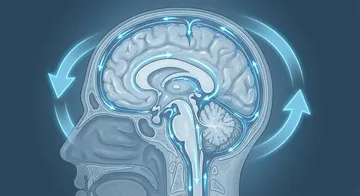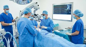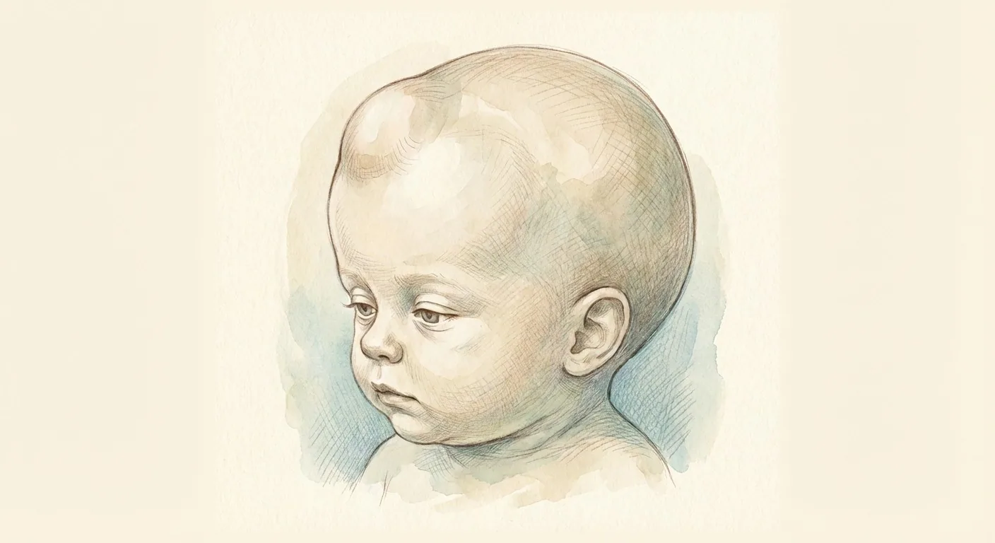
Hydrocephalus is one of the most common neurosurgical pathologies encountered in the neonatal period. Because the fontanelles are open, infants present with an increase in head circumference — presenting a different clinical picture from adults.

Causes
Congenital: Aqueductal stenosis, hydrocephalus seen together with myelomeningocele (open spine), and Dandy-Walker malformation.
Acquired: Intraventricular haemorrhage (most common in premature infants), post-meningitis, tumour-related obstruction.1,2
Symptoms
Infants (open fontanelle)
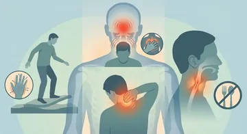
Rapid increase in head circumference (crossing the percentile curve), tense and bulging fontanelle, setting sun sign (eyes looking downward), irritability, feeding difficulties, vomiting, developmental delay.
Older Children (closed fontanelle)
Headache (especially in the morning), nausea-vomiting, blurred vision (papilledema), decline in school performance, personality changes, gait disturbance.
Diagnosis
Prenatal Ultrasound: Ventricular enlargement can be detected in the 18-20th week ultrasound.3
Postnatal Ultrasound: Performed through the open fontanelle, ventricular sizes are monitored. It is the first-choice imaging.
MRI: The gold standard for anatomical detail, evaluating the cause of hydrocephalus, and accompanying anomalies.4
Head Circumference Monitoring: It should be measured at every follow-up examination and tracked on the percentile curve.
Treatment
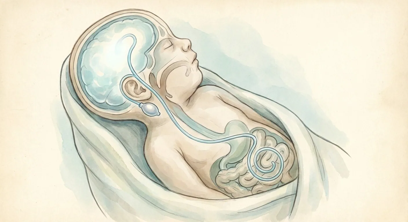
VP Shunt: The most commonly performed surgical treatment.5,6 Low-profile valves specific to infants are used. Shunt revision may be required with growth.
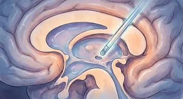
ETV ± Choroid Plexus Coagulation: It may be preferred to increase the success rate in the treatment of obstructive hydrocephalus in infants.7,8 Choroid plexus coagulation reduces CSF production.
Temporary Measures: Serial lumbar puncture (posthemorrhagic hydrocephalus), external ventricular drainage (EVD), subgaleal shunt.
In obstructive hydrocephalus in infants, choroid plexus coagulation along with ETV may be preferred to increase the success rate.
→ Next page: S50 — Chiari Malformation
Kaynaklar
- Darko K et al. Childs Nerv Syst. 2026. PMID 42371150
- Valdez Sandoval P et al. Childs Nerv Syst. 2019. PMID 30953157
- Loo CK et al. Pathology. 2004. PMID 15203729
- Roth J et al. Pediatr Neurosurg. 2019. PMID 30870836
- Fagundes W et al. World Neurosurg. 2025. PMID 40545034
- Elmesallamy WAEA et al. Childs Nerv Syst. 2023. PMID 36550332
- Yadav YR et al. Asian J Neurosurg. 2016. PMID 27695532
- Zhu X et al. Childs Nerv Syst. 2013. PMID 23151740
Doç. Dr. Özgür Akşan
Beyin ve Sinir Cerrahisi Uzmanı
Beyin ve Sinir · Sorumlu Yazı İşleri Müdürü ve Editör · Künye