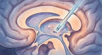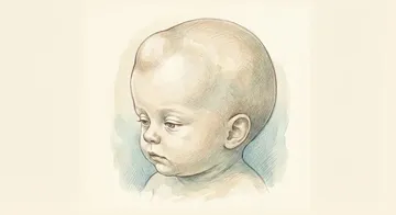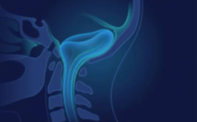
Symptomatic Chiari Type I (especially progressive neurological deficit), CSF flow disruption on MRI, and the presence of syringomyelia (especially an expanding syrinx) are the main indications for decompression surgery.
Posterior Fossa Decompression
Standard Technique
With a 5-6 cm incision in the middle of the back of the neck, the lower part of the occipital bone and the posterior arch of C1 (atlas) are removed. With this bony decompression, the foramen magnum is widened, providing more space for the cerebellar tonsils.
With a 5-6 cm incision in the middle of the back of the neck, the lower part of the occipital bone and the posterior arch of C1 (atlas) are removed.
Opening the Dura (Duraplasty)
Most surgeons open the dura and close it with an expansile patch (duraplasty); however, evidence that this method restores CSF circulation more effectively than decompression without duraplasty is conflicting.1,2 Autologous pericranium, artificial dura materials, or pericardial grafts can be used.
Tonsillectomy/Tonsillar Cauterisation
The herniated tonsils are shrunk or lifted to open the CSF flow pathway. Aggressive tonsillar resection is controversial.
Is Bony Decompression Sufficient?
The "bone-only" approach is preferred in some centres. Its advantage: the risk of CSF leak is very low. Its disadvantage: it can lead to inadequate decompression in some patients.3,4 CSF flow study can be checked with ultrasound during surgery.
Syringomyelia
Pathophysiology
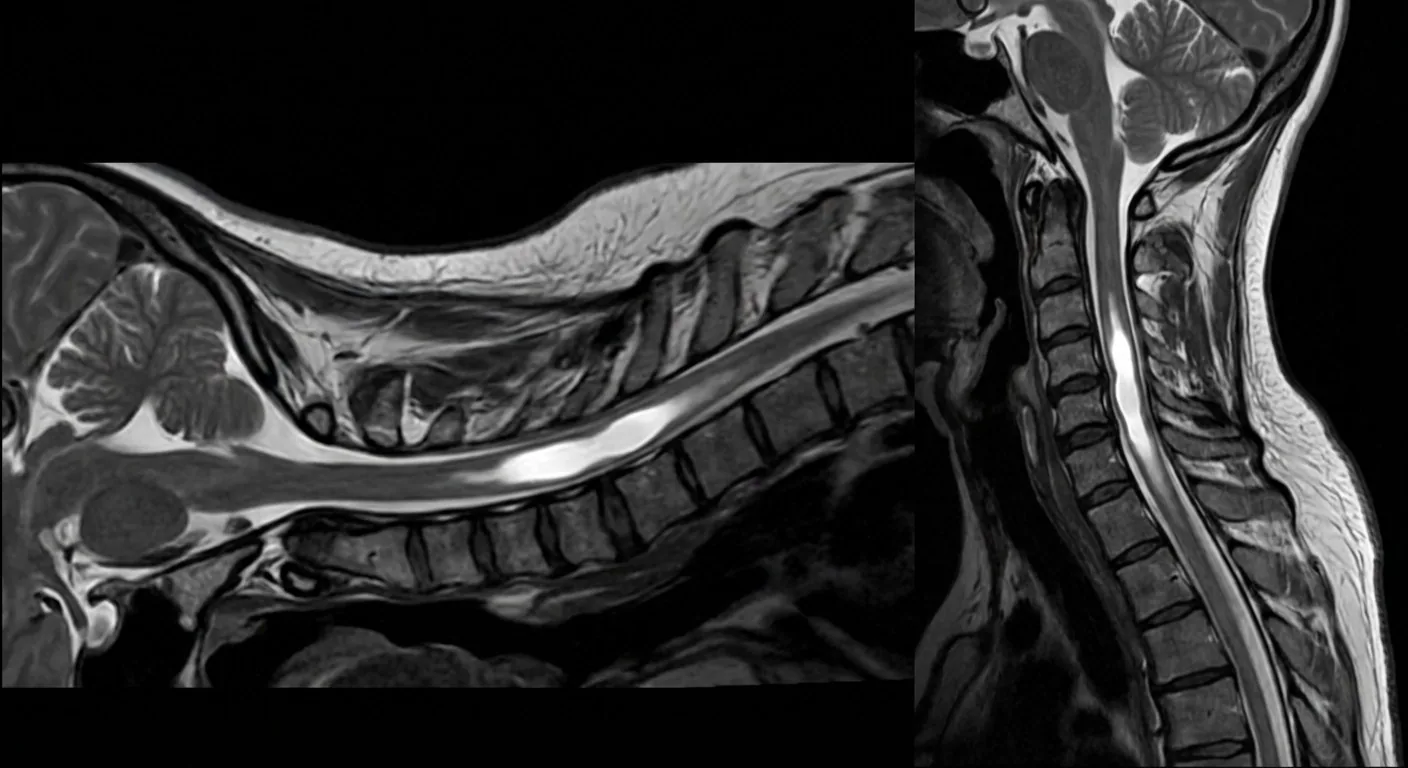
In Chiari, the obstruction of CSF flow at the level of the foramen magnum leads to fluid accumulation (syrinx) within the spinal cord. The syrinx compresses the spinal cord from the inside, causing neurological deficits.
Treatment
If Chiari decompression is successful, the syrinx shrinks in most cases; this rate has been reported as 80-90% in series.5,6 In resistant syrinx: a syringo-subarachnoid shunt or syrinx drainage is considered.
Follow-up
An MRI check is performed in the 3rd month after surgery, and subsequent MRI follow-ups are generally repeated at 12 to 18 months.7 In patients with a syrinx, avoiding Valsalva manoeuvres (heavy lifting, straining) is recommended.
Outcomes
Improvement in headache after decompression has been reported in the literature at approximately 79 percent, and up to 98 percent in the series of experienced centres.8 Neurological deficits show a 50-70 percent improvement.9 Syringomyelia shrinks or remains stable in most cases.
→ Next page: S52 — Brain Haemorrhage and Stroke

Kaynaklar
- Tam SKP et al. Acta Neurochir (Wien). 2021. PMID 32577895
- Yuan C et al. Turk Neurosurg. 2022. PMID 35652180
- Durham SR et al. J Neurosurg Pediatr. 2008. PMID 18590394
- Lin W et al. World Neurosurg. 2018. PMID 29138073
- Alzain A et al. Cureus. 2025. PMID 41541935
- Turk O et al. Med Sci Monit. 2025. PMID 39799391
- Deng K et al. Chin Med J (Engl). 2010. PMID 20819571
- Arnautovic KI et al. World Neurosurg. 2021. PMID 33249219
- Saez RJ et al. J Neurosurg. 1976. PMID 956878
Doç. Dr. Özgür Akşan
Beyin ve Sinir Cerrahisi Uzmanı
Beyin ve Sinir · Sorumlu Yazı İşleri Müdürü ve Editör · Künye