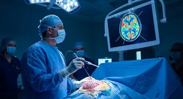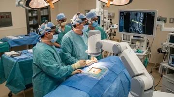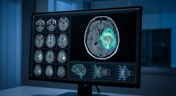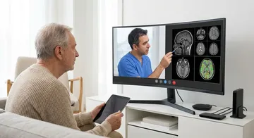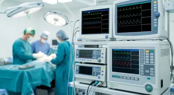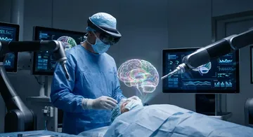
In special rooms equipped with an MRI scanner inside the operating room, an MRI can be taken during the surgery. This allows the tumour removal rate to be evaluated instantly.
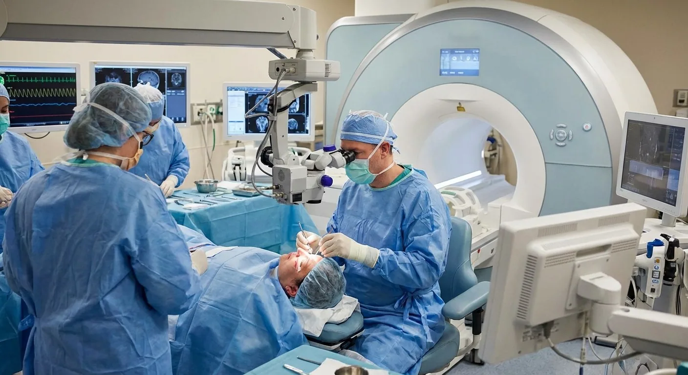
How Does It Work? After tumour removal, the patient is taken to the MRI scanner on the operating table. If there is a residual (remaining) tumour, additional surgery is performed immediately. The tumour is completely removed without waking the patient.
Advantages: Although advanced imaging methods like intraoperative MRI increase the gross total resection rate by approximately 16%, this difference was not found to be statistically significant.1 It reduces the need for a second surgery. It solves the brain shift problem (navigation with updated images).
Fluorescence-Guided Surgery
5-ALA (5-Aminolevulinic Acid)
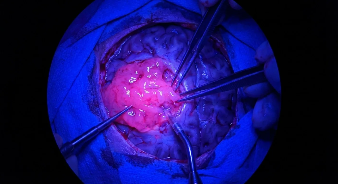
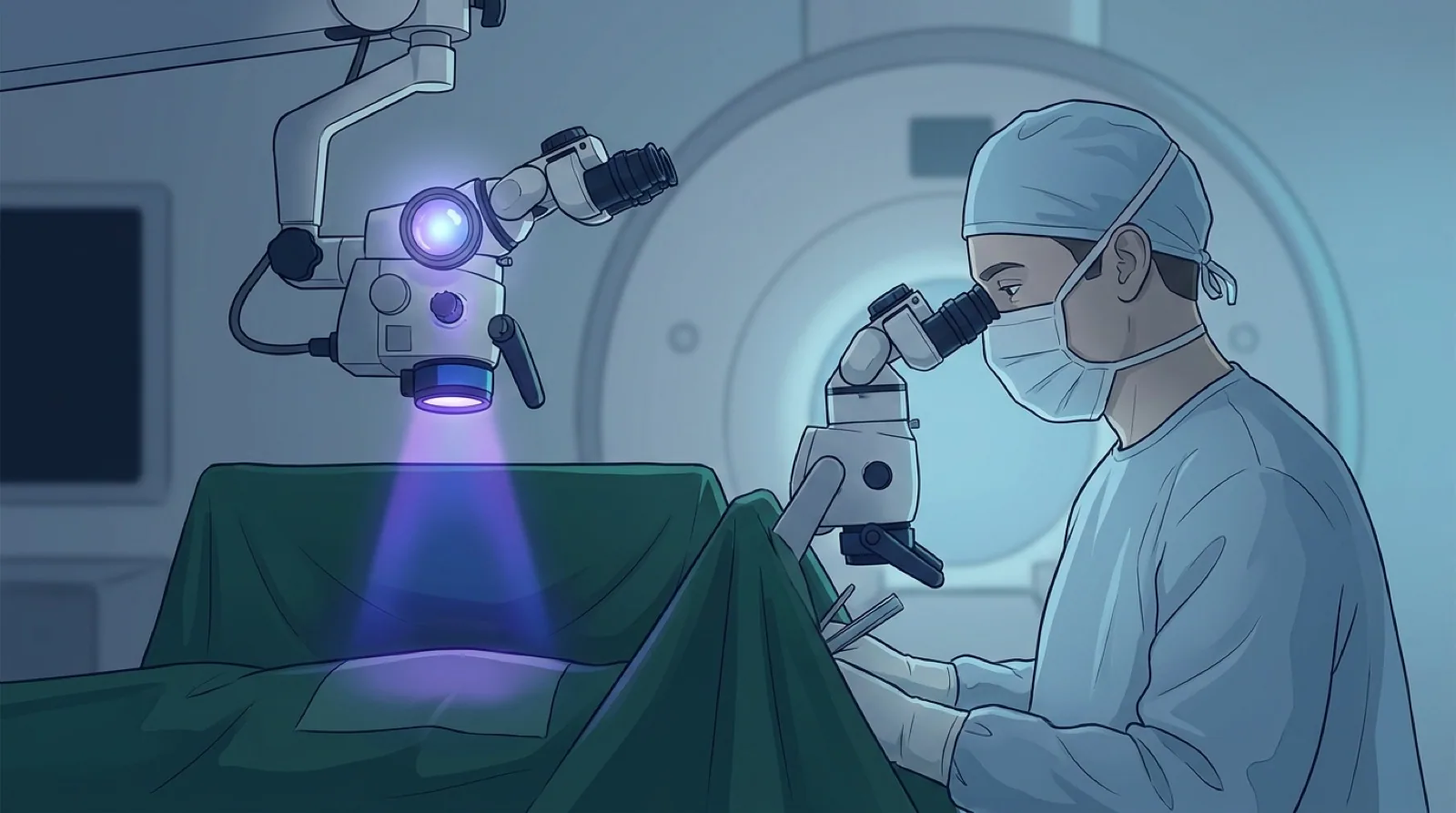
Patients are given oral 5-ALA 3 hours before surgery. Tumour cells convert this substance into protoporphyrin IX. When the surgical microscope is switched to a blue filter, the tumour tissue glows pink-purple, while normal tissue does not glow.
The use of 5-ALA significantly increases the gross total resection rate in glioblastoma.2,3 It is most effective for WHO grade 3-4 gliomas.4,5
Fluorescein Sodium (FNa)
A fluorescent dye administered intravenously. It accumulates in areas where the blood-brain barrier is disrupted (tumour) and appears yellow-green. It is cheaper than 5-ALA and has a better side effect profile.6
Indocyanine Green (ICG)
A near-infrared fluorescent dye. It shows vascular flow in real time. It is used in aneurysm surgery to verify flow after clipping and in AVM surgery to identify feeding and draining arteries.
→ Next page: S65 — 3D Printing and Custom-Made Implants
Kaynaklar
- Caras A et al. World Neurosurg. 2020. PMID 31639502
- Eljamel S. Int J Mol Sci. 2015. PMID 25961952
- Mueed A et al. Neurosurg Rev. 2026. PMID 42493686
- Eatz TA et al. J Neurooncol. 2022. PMID 34989964
- Almekkawi AK et al. Oper Neurosurg. 2020. PMID 31828346
- Smith EJ et al. World Neurosurg. 2021. PMID 34492388
- Sanikommu S et al. Neurosurg Rev. 2026. PMID 42020844
Doç. Dr. Özgür Akşan
Beyin ve Sinir Cerrahisi Uzmanı
Beyin ve Sinir · Sorumlu Yazı İşleri Müdürü ve Editör · Künye