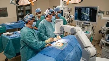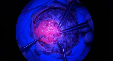
Augmented reality is the technology that superimposes digital information onto a real-world view. Through special glasses or microscope integration, the surgeon can see the patient's anatomy, tumour margins, and critical structures projected directly onto the surgical field.

How Does It Work?
Preoperative MRI/CT images are converted into a 3D model. The AR system aligns this model with the patient's actual anatomy (registration). Through their glasses or microscope, the surgeon sees both the real surgical field and the superimposed digital information.
Application Areas
Incision Planning: The most appropriate craniotomy site is determined by projecting the tumour onto the skin.1,2
Visualisation of Tumour Margins: During surgery, tumour margins, eloquent areas, and blood vessels are displayed holographically.
Spine Surgery: Projecting the pedicle screw trajectory onto the spine. It reduces radiation exposure.3,4
Education: Young surgeons can learn by sharing the AR view seen by the experienced surgeon.
Low-latency AR streaming with 5G, automatic anatomical structure recognition with AI, haptic (tactile) feedback, and AR + robotic surgery integration are promising developments.
Current Systems
Devices such as Microsoft HoloLens, Apple Vision Pro, and Magic Leap are being adapted for surgical applications. Zeiss and Leica microscope-integrated AR systems are ready for clinical use.
The Future
Low-latency AR streaming with 5G, automatic anatomical structure recognition with AI, haptic (tactile) feedback, and AR + robotic surgery integration are promising developments.
→ Next page: S71 — Protect Your Brain
Kaynaklar
- Contreras López WO et al. Clin Neurol Neurosurg. 2019. PMID 30579049
- Van Gestel F et al. Front Neurol. 2023. PMID 36998774
- McCloskey K et al. World Neurosurg. 2023. PMID 36812986
- Edström E et al. Spine (Phila Pa 1976). 2020. PMID 31415457

Doç. Dr. Özgür Akşan
Beyin ve Sinir Cerrahisi Uzmanı
Beyin ve Sinir · Sorumlu Yazı İşleri Müdürü ve Editör · Künye