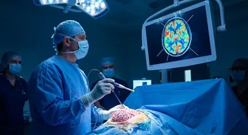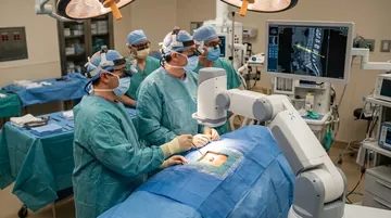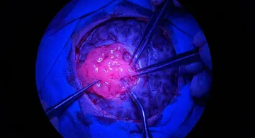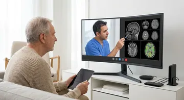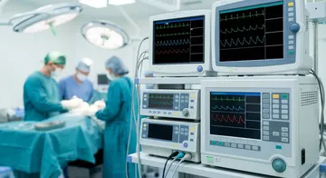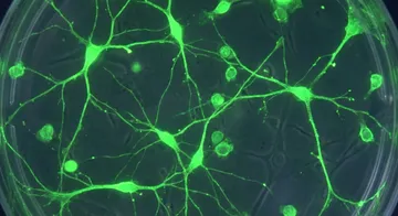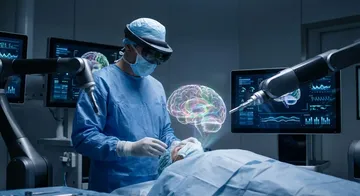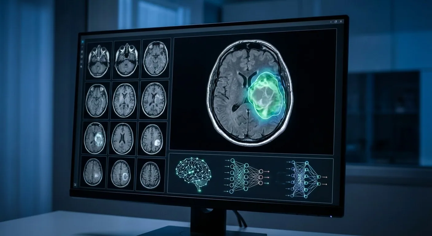
Artificial intelligence algorithms provide decision support to radiologists and surgeons by analysing MRI, CT, and PET images. Deep learning models learn from millions of images to classify tissues, detect anomalies, and make measurements.
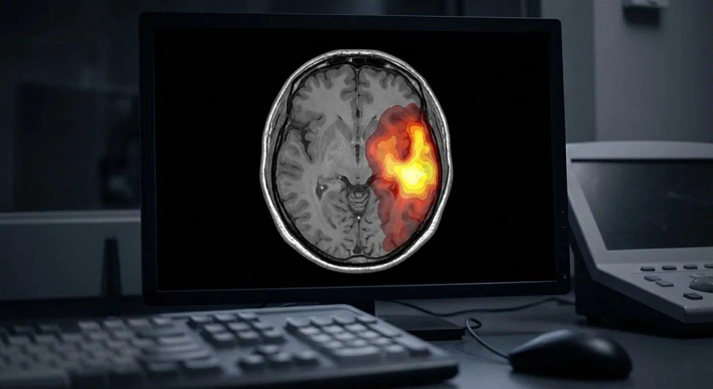
Application Areas
Tumour Segmentation: AI automatically draws tumour boundaries and calculates their volume in MRI images. Tumour segmentation with artificial intelligence accelerates surgical planning and treatment response monitoring processes.1,2
Stroke Detection: Artificial intelligence optimises the workflow by supporting the rapid detection of haemorrhage or ischaemia in CT images.3,4 It enables the preliminary evaluation of emergency cases with "AI triage".
Degenerative Diseases: Hippocampal volume measurement for Alzheimer's, substantia nigra analysis for Parkinson's. It provides the opportunity for early diagnosis.5,6
Spine Analysis: Disc degeneration grading, stenosis measurement, Cobb angle calculation. It shortens the reporting time.7,8
The Situation in Turkey
Many hospitals in Turkey have started using AI-supported imaging solutions. FDA and CE-approved AI software are taking their place in daily practice. However, AI results must always be verified by a specialist doctor — AI is not a decision-maker, but a decision supporter.9
Artificial intelligence helps in the evaluation by analysing the hippocampus volume in Alzheimer's and the substantia nigra in Parkinson's.
Ethics and Security
Transparency (explainability) is important in AI imaging analysis. The guidance of clinical decisions by "black box" algorithms raises ethical questions. Patient data security, KVKK compliance, and false positive/negative rates must be carefully evaluated.
→ Next page: S67 — Telemedicine
Kaynaklar
- Qu Y et al. Med Image Anal. 2021. PMID 33550006
- Sanker V et al. Front Oncol. 2025. PMID 41626168
- Ha SH et al. Clin Exp Emerg Med. 2026. PMID 41554281
- Hu P et al. Int J Surg. 2024. PMID 38489547
- Wang LX et al. Arq Neuropsiquiatr. 2024. PMID 39146974
- Reddy S et al. Cureus. 2024. PMID 38784299
- Rajmohamed RF et al. Acad Radiol. 2025. PMID 41015710
- Lim DSW et al. Radiology. 2022. PMID 35699577
- Bergquist M et al. Eur Radiol. 2024. PMID 37505245
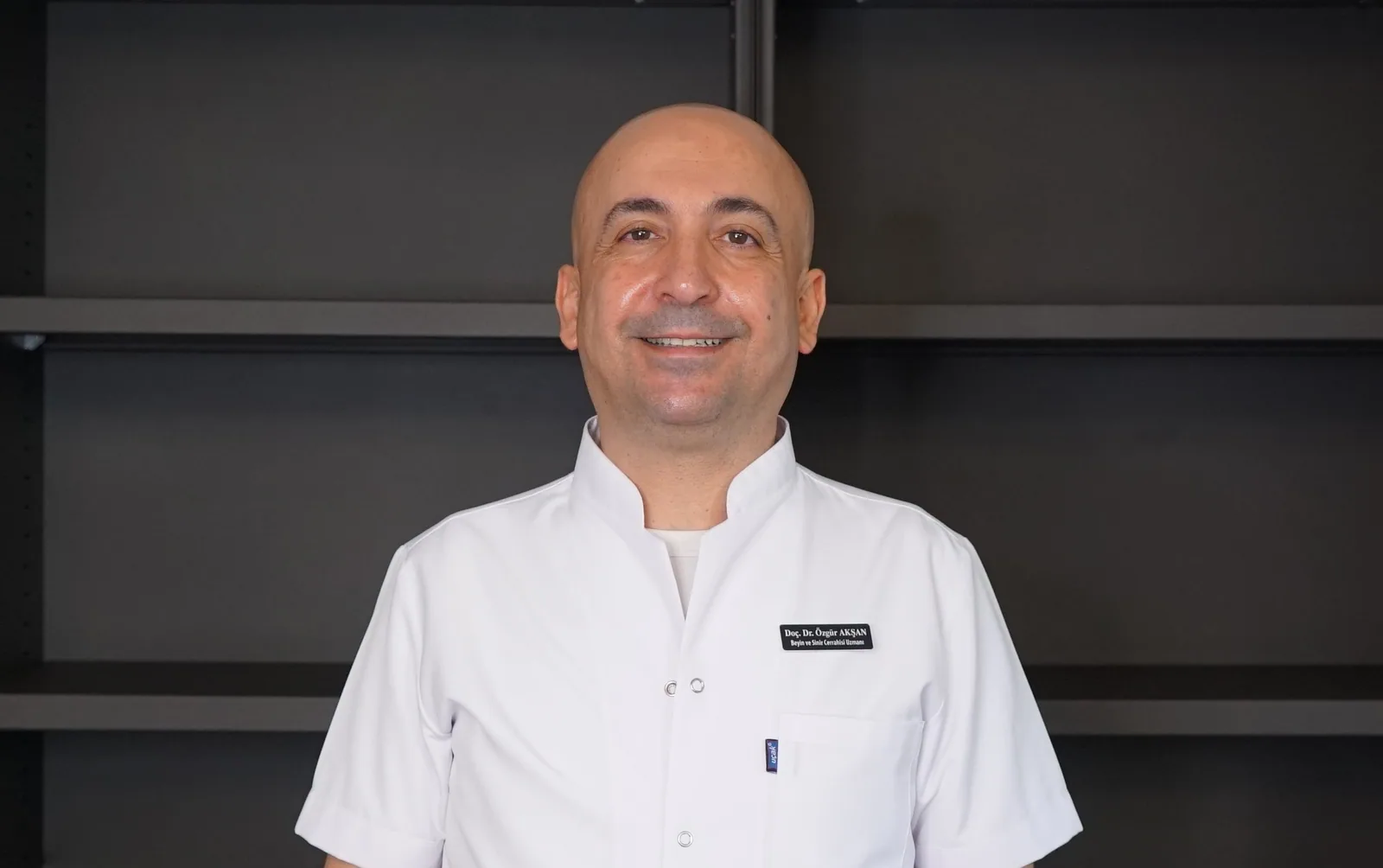
Doç. Dr. Özgür Akşan
Beyin ve Sinir Cerrahisi Uzmanı
Beyin ve Sinir · Sorumlu Yazı İşleri Müdürü ve Editör · Künye