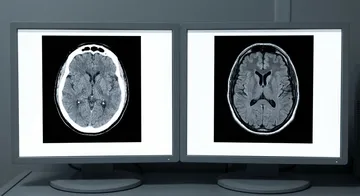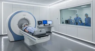
In modern neurosurgery, the goal is not only to remove the tumour but to preserve the patient's speech, movement, vision, and cognitive functions while doing so. Functional MRI (fMRI) and diffusion tensor imaging (DTI) are indispensable tools in achieving this balance.
What is Functional MRI (fMRI)?
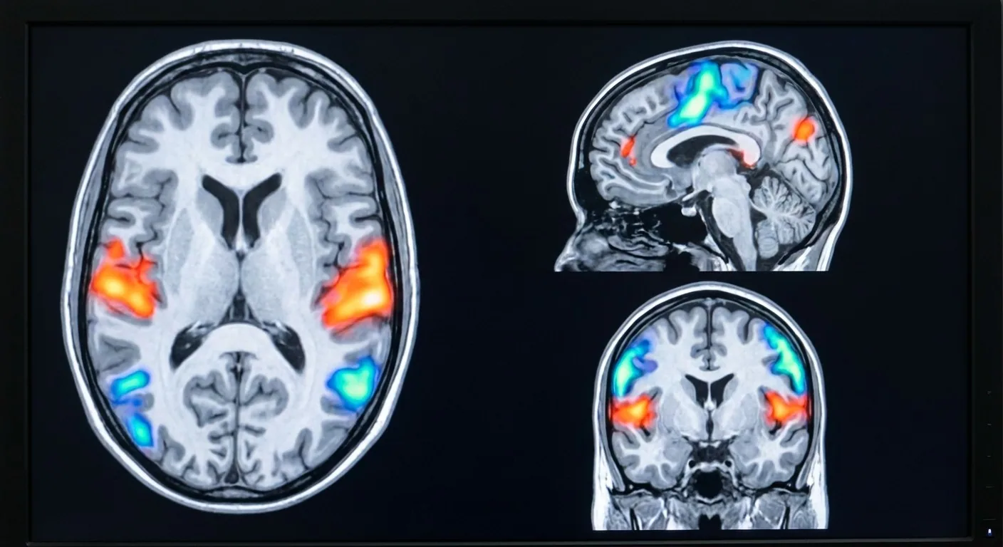
Functional MRI is an imaging method that shows which regions of the brain become active during specific tasks. While inside the scanner, the patient performs finger movements, generates words, or looks at visual stimuli; during this time, the activated brain regions are marked on a map.
Thanks to this mapping, the surgeon knows the distance of the tumour to critical regions such as the motor cortex, speech centres (Broca's and Wernicke's areas), or the visual cortex before the surgery. This information plays a fundamental role in planning the surgical approach and informing the patient about the risks.
Diffusion Tensor Imaging (DTI) and Tractography
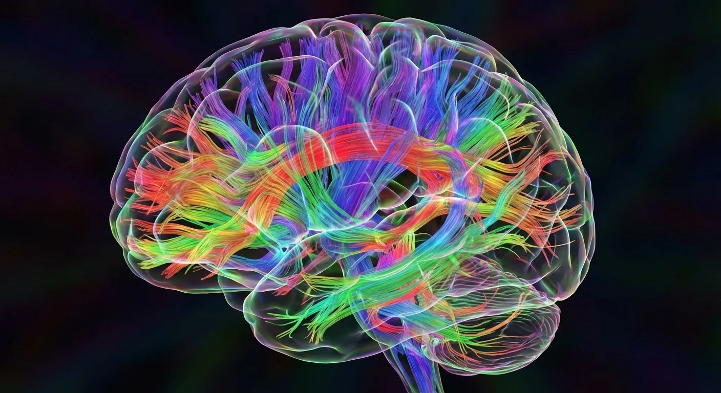
DTI is an MRI technique that shows the direction and integrity of nerve fibres (white matter tracts) within the brain. Tractography, on the other hand, is a three-dimensional nerve fibre map created from DTI data.
Thanks to tractography, the distance of critical nerve bundles such as the corticospinal tract (movement pathway), arcuate fasciculus (speech pathway), and optic radiation (vision pathway) to the tumour, as well as their displacement or infiltration by the tumour, are evaluated. This information allows the surgeon to determine safe resection margins.
This information plays a fundamental role in planning the surgical approach and informing the patient about the risks.
MR Spectroscopy
MR spectroscopy measures the biochemical components in the tumour region. N-acetyl aspartate (NAA), choline, creatine, and lactate levels provide clues about the grade and aggressiveness of the tumour. A high choline and low NAA ratio suggests a high-grade tumour.1,2 This technique helps to make predictions about the tumour before a biopsy is performed.
Integration in Surgical Planning
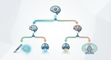
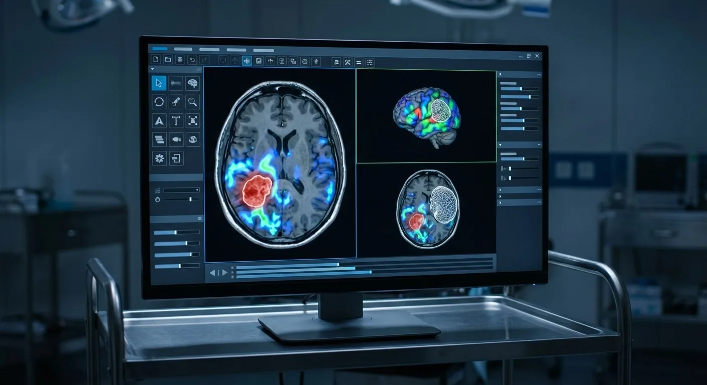
In modern neurosurgery practice, all these data — structural MRI, fMRI, DTI/tractography, and spectroscopy — are integrated into the neuronavigation system. During the surgery, the surgeon proceeds by seeing these data in real-time on the screen.
→ Next page: S09 — Glioblastoma (GBM)
Kaynaklar
- Wang Q et al. Eur Radiol. 2016. PMID 26471274
- Usinskiene J et al. Neuroradiology. 2016. PMID 26767528
- Lo YT et al. World Neurosurg. 2021. PMID 33540099
- Li Z et al. Eur J Cancer. 2024. PMID 38218157
Doç. Dr. Özgür Akşan
Beyin ve Sinir Cerrahisi Uzmanı
Beyin ve Sinir · Sorumlu Yazı İşleri Müdürü ve Editör · Künye