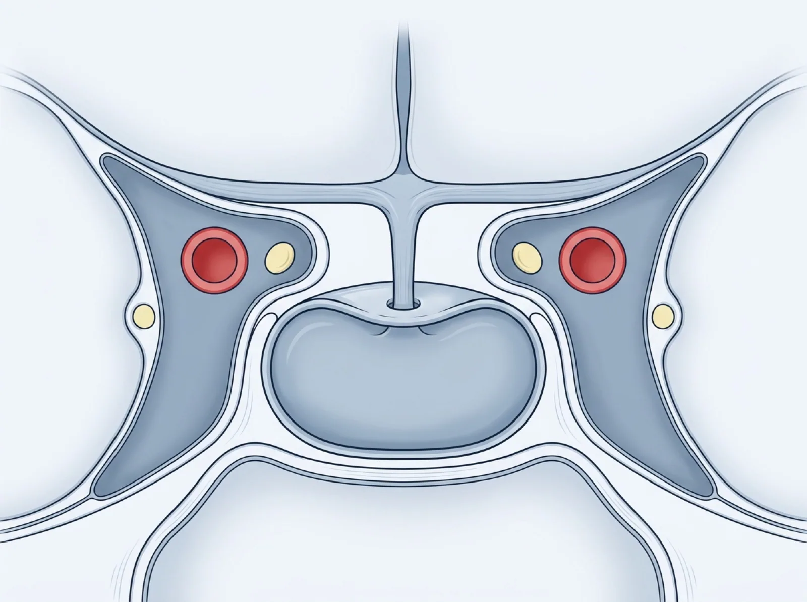
The pituitary gland is an endocrine organ, approximately the size of a chickpea, located at the base of the brain within a bony structure called the Turkish saddle (sella turcica). It plays a central role in regulating many hormones in the body.
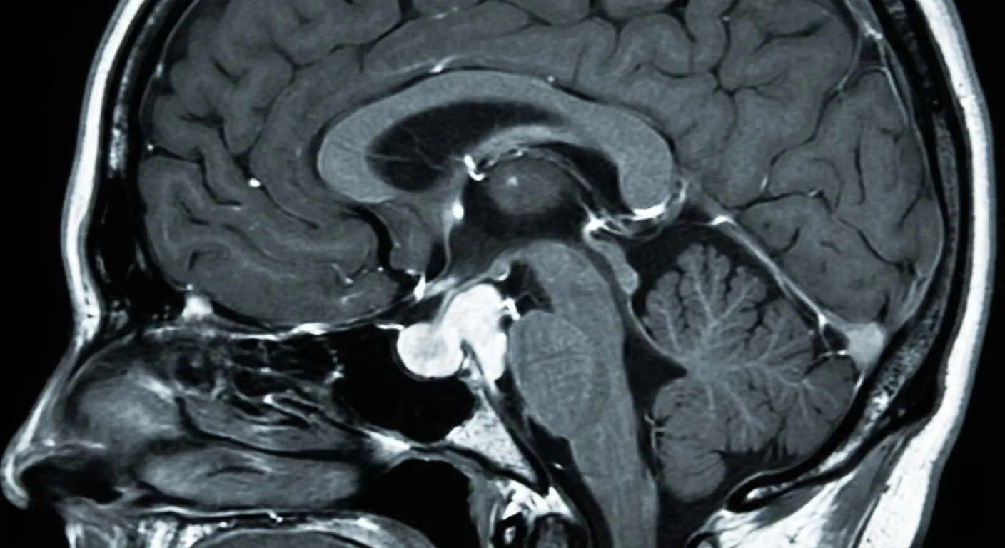
What is a Pituitary Adenoma?
Pituitary adenomas are almost always benign. They are divided into two according to their size: microadenomas (under 10 mm) and macroadenomas (10 mm and over). Functionally, they are classified as hormone-secreting (functional) and non-hormone-secreting (non-functional) adenomas.
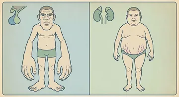
Functional adenomas lead to distinct clinical pictures due to excessive hormone production. Prolactinoma is the most common functional adenoma; it manifests with menstrual irregularity and milk discharge in women, and sexual dysfunction in men. Growth hormone-secreting adenomas cause acromegaly or gigantism, while ACTH-secreting ones cause Cushing's disease.
Non-functional adenomas, on the other hand, cause symptoms when they grow and compress surrounding structures. They most commonly present with visual impairment (especially bitemporal hemianopsia — loss of visual field on both sides) and headache.
Diagnosis
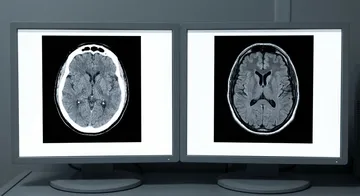
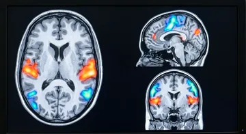
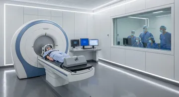
Hormonal evaluation and imaging are performed together. In blood tests, prolactin, growth hormone, IGF-1, cortisol, ACTH, thyroid hormones, and gonadotropins are measured. Sellar MRI (thin-slice, with contrast) shows the size of the tumour, cavernous sinus invasion, and its relationship to the optic chiasm.
Transsphenoidal Endoscopic Surgery
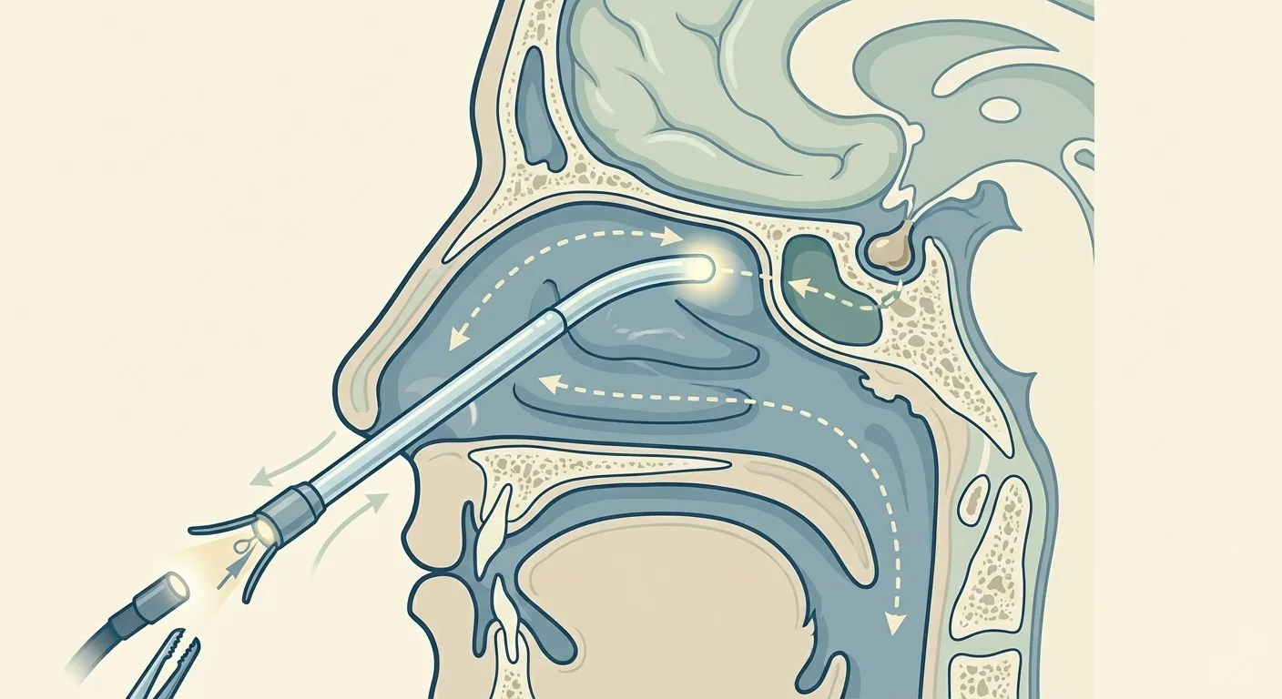
The gold standard of modern pituitary surgery is the endoscopic transsphenoidal approach.1,2 In this technique, the pituitary gland is reached through the nostril, via the sphenoid sinus. The skull is not opened, and brain tissue is not manipulated.
Endoscopic imaging provides a wide field of view and high resolution, allowing the surgeon to remove the tumour more safely and comprehensively. The hospital stay in endoscopic transsphenoidal surgery is generally 2-3 days.3,4
Surgical indications include macroadenomas causing vision loss, functional adenomas resistant to medical treatment (excluding prolactinoma), and apoplexy (pituitary haemorrhage). In prolactinoma, the first treatment is usually medical (dopamine agonists), and surgery is considered in cases of drug resistance or intolerance.5,6
Post-Surgery
Postoperative follow-up is done with hormonal evaluation, visual field testing, and control MRI. Diabetes insipidus (temporary water imbalance) is the most common early complication; it is mostly temporary. CSF fistula (cerebrospinal fluid leak) is a rare but important complication.
→ Next page: S17 — Hormonal Evaluation in Pituitary Tumours
Kaynaklar
- Zaletel T et al. Pituitary. 2026. PMID 41604016
- Sadhwani N et al. World Neurosurg. 2024. PMID 38734175
- Saad M et al. J Clin Med. 2022. PMID 36233696
- Vimawala S et al. Int Forum Allergy Rhinol. 2020. PMID 32362064
- Cai X et al. Chin Neurosurg J. 2022. PMID 35395837
- Ottenhausen M et al. Neurosurg Rev. 2023. PMID 37249700
- Yi N et al. Endocrine. 2018. PMID 29934876
- Zhen JR et al. Zhonghua Fu Chan Ke Za Zhi. 2008. PMID 18843964
Doç. Dr. Özgür Akşan
Beyin ve Sinir Cerrahisi Uzmanı
Beyin ve Sinir · Sorumlu Yazı İşleri Müdürü ve Editör · Künye