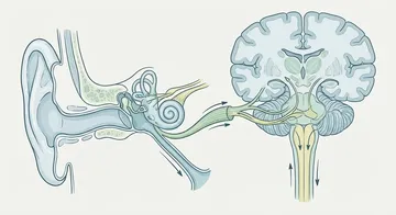
When a brain tumour is suspected, the diagnostic process begins with a neurological examination and becomes clear with imaging methods. Making the correct diagnosis forms the foundation of the treatment plan.
Neurological Examination
Everything begins with a comprehensive neurological examination. The doctor systematically evaluates muscle strength, reflexes, sensation, balance, coordination, visual fields, and cognitive functions. This examination helps predict the location of the possible lesion and determines which imaging method is appropriate.
Brain MRI (Magnetic Resonance Imaging)
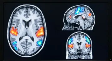
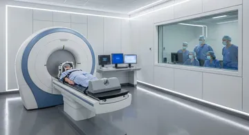
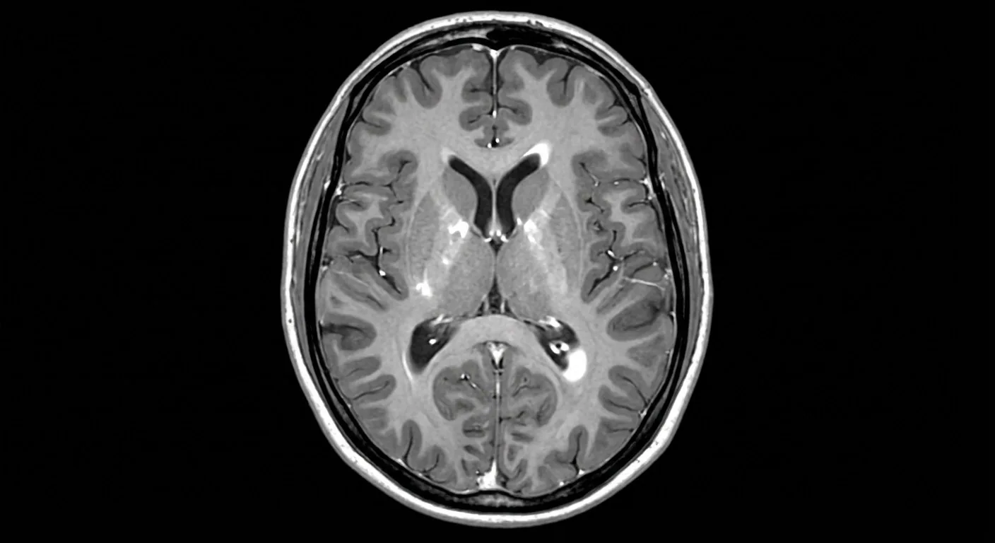
Thanks to its soft tissue contrast, MRI allows for the estimation of the tumour's size, location, relationship with surrounding tissues, and in some cases, its histological type.
Standard MRI sequences include T1-weighted (without and with contrast), T2-weighted, FLAIR, and diffusion-weighted imaging. In contrast-enhanced imaging, a gadolinium-based contrast agent is used; the areas where the tumour disrupts the blood-brain barrier appear bright.
Standard MRI sequences include T1-weighted (without and with contrast), T2-weighted, FLAIR, and diffusion-weighted imaging.
Computed Tomography (CT)
CT is the first-choice imaging method, especially in emergency situations. Its rapid acquisition, widespread availability, and good visualisation of bone structures are its advantages. However, it falls behind MRI in soft tissue differentiation. CT shows bleeding, calcification, and bone invasion well.1
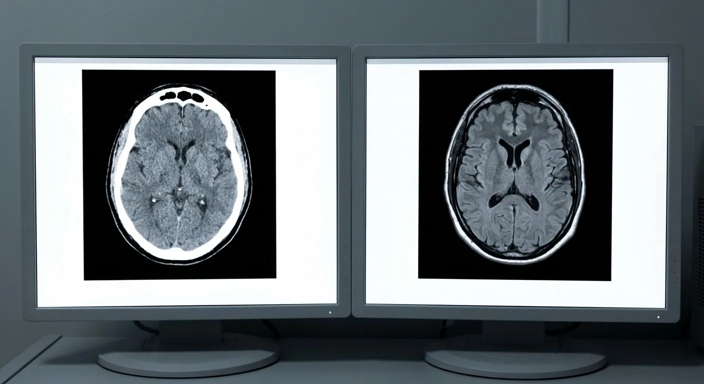
Comparison of MRI and CT
MRI is superior in soft tissue detail, does not involve radiation, and can obtain multi-planar slices. CT, on the other hand, is faster, shows bone pathologies better, and is preferred in emergencies. In most patients, both methods are used together.
Advanced Imaging Techniques
MR spectroscopy helps predict the tumour type by analysing the chemical composition of the tumour tissue.2,3
These advanced techniques will be discussed in detail on the next page.
→ Next page: S08 — Functional MRI and Tractography
Kaynaklar
- Wietelmann D et al. Radiologe. 1998. PMID 9861650
- Wang W et al. PLoS One. 2014. PMID 25393009
- Wang Q et al. Tumour Biol. 2017. PMID 28631566
- Behzadi AH et al. Radiology. 2018. PMID 28846495
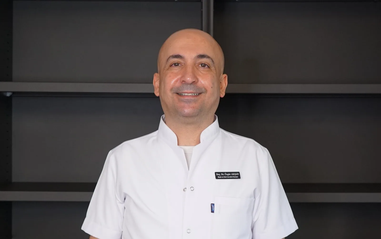
Doç. Dr. Özgür Akşan
Beyin ve Sinir Cerrahisi Uzmanı
Beyin ve Sinir · Sorumlu Yazı İşleri Müdürü ve Editör · Künye