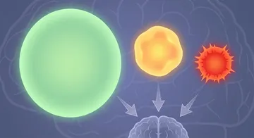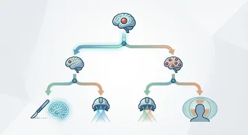
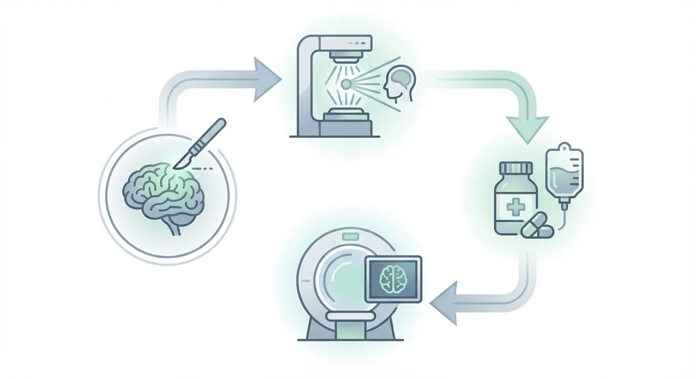
GBM treatment does not consist of a single stage; it is a structured process in which surgery, radiotherapy, and chemotherapy are applied in a specific sequence and timing. This process is called the Stupp Protocol and is the worldwide standard approach for GBM treatment.1,2
Treatment Steps
The first step is surgical resection. After maximum safe resection is performed, the definitive diagnosis, molecular subtype, and prognostic markers are evaluated through pathological examination. Molecular markers such as MGMT methylation status, IDH mutation, and 1p/19q co-deletion play a critical role in predicting treatment response and prognosis.
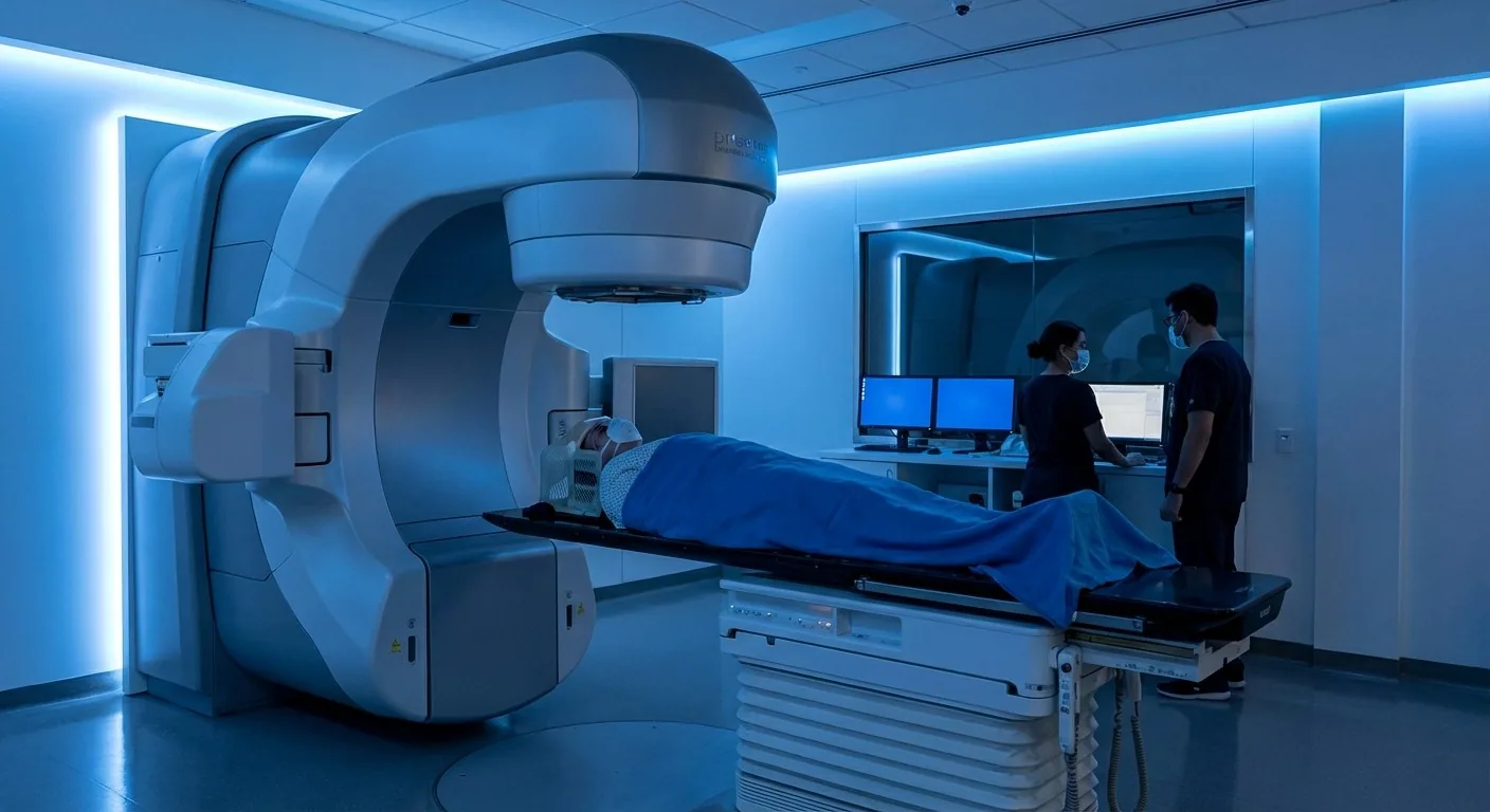
The second step is concurrent chemoradiotherapy. Radiotherapy is started approximately 4-6 weeks after surgery. The standard application is given at a total dose of 60 Gy, in 30 fractions, over 6 weeks.3,4 During this period, daily temozolomide (TMZ) chemotherapy is administered concurrently.
The third step is adjuvant chemotherapy. Following a 4-week rest period after the completion of chemoradiotherapy, 6 cycles of temozolomide chemotherapy are administered for 5 days a month; in selected patients, the treatment can be extended up to 12 cycles.5,6
The Importance of Molecular Markers
MGMT promoter methylation is one of the most important prognostic markers in GBM. In patients with methylated MGMT, the response to temozolomide treatment is significantly better.7,8 Therefore, molecular analysis is a part of the standard practice in post-surgical pathological evaluation.
The presence of an IDH mutation is associated with a better prognosis. Grade 4 astrocytomas carrying an IDH mutation show longer survival compared to IDH wild-type glioblastoma.9,10
Although MRI follow-up is generally recommended every 3 months or every 3-6 months according to guidelines, in some clinical practices, an MRI is performed every 3 months for the first year, and every 6 months thereafter.
Follow-Up Process
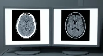
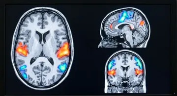
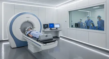
During and after treatment, follow-up is performed with regular contrast-enhanced brain MRIs. Although MRI follow-up is generally recommended every 3 months or every 3-6 months according to guidelines, in some clinical practices, an MRI is performed every 3 months for the first year, and every 6 months thereafter.11,12 Distinguishing between pseudoprogression (a temporary appearance of growth due to treatment) and true progression is important during follow-up.
In Case of Recurrence
Unfortunately, recurrence is an expected situation in GBM. When recurrence develops, a second surgery, bevacizumab treatment, different chemotherapy regimens, or clinical trial protocols can be evaluated. Tumour treating fields (TTFields) technology is also an innovative approach used in recurrent and newly diagnosed GBM.13,14
→ Next page: S11 — Current Research in GBM
Kaynaklar
- McBain C et al. Cochrane Database Syst Rev. 2021. PMID 34559423
- Calvin N et al. Surg Neurol Int. 2024. PMID 39640319
- Stupp R et al. N Engl J Med. 2005. PMID 15758009
- Mallick S et al. J Neurooncol. 2023. PMID 37452916
- Attarian F et al. Front Oncol. 2021. PMID 34900732
- Fasano M et al. Oncol Lett. 2024. PMID 39006948
- Feldheim J et al. Cancers (Basel). 2019. PMID 31766430
- Buonaiuto M et al. J Transl Med. 2025. PMID 40640872
- Chen JR et al. Medicine (Baltimore). 2016. PMID 26945349
- Sarac ME et al. J Clin Med. 2025. PMID 40217970
- Anvari K et al. Front Oncol. 2024. PMID 38774410
- INTERVAL-GB Collaborative et al. J Neurooncol. 2024. PMID 39105956
- Khagi S et al. Oncologist. 2025. PMID 39401002
- Ornelas AS et al. Neurologist. 2019. PMID 30817495
Doç. Dr. Özgür Akşan
Beyin ve Sinir Cerrahisi Uzmanı
Beyin ve Sinir · Sorumlu Yazı İşleri Müdürü ve Editör · Künye