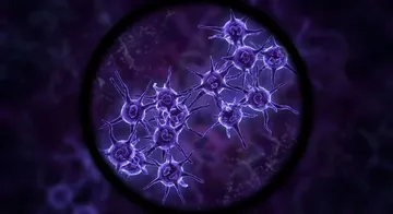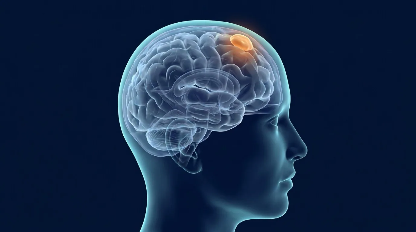
Meningioma is the most common benign intracranial tumour originating from the brain membranes (meninges). It accounts for approximately 35-40 percent of primary brain tumours.1,2 It is seen twice as often in women as in men, and its incidence increases with age.3,4
What is a Meningioma?
Meningiomas originate from the cells in the arachnoid layer of the brain membrane. They grow outside the brain, on the membranes, and cause symptoms by pressing on the brain tissue. Most grow slowly; some meningiomas can remain stable for years without growing.
Locations
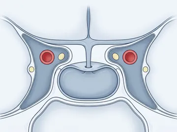
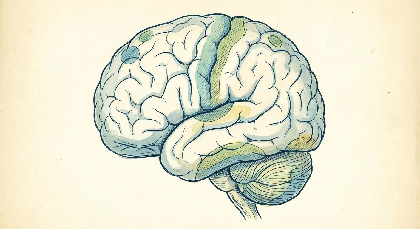
Meningiomas can be seen in many different locations within the skull. The most common locations are the convexity (upper surface of the brain), parasagittal region (midline), sphenoid wing, tuberculum sellae, olfactory groove, posterior fossa, and foramen magnum. The location directly determines the symptoms and the surgical approach.
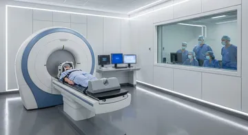
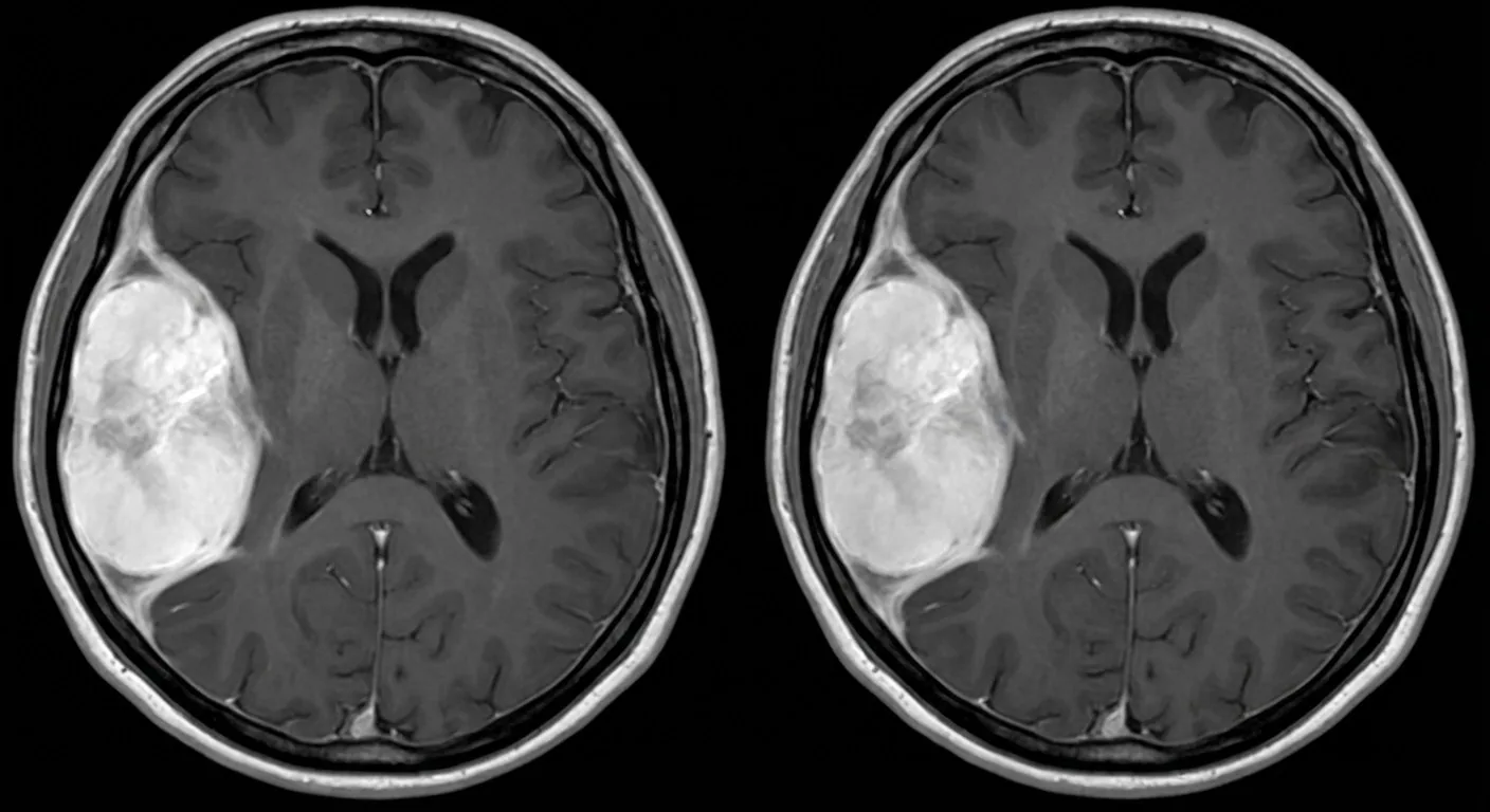
On contrast-enhanced brain MRI, meningiomas are typically observed as homogeneous, intensely contrast-enhancing masses showing a broad-based contact with the dura.
Symptoms
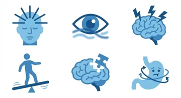
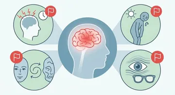
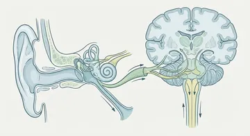
Headache is the most common symptom. Besides this, seizures, vision loss (in those pressing on the optic nerve), loss of smell (in olfactory groove meningiomas), hearing loss and balance disorders (in posterior fossa meningiomas), and loss of strength in the arm or leg (in those near the motor cortex) can be seen. Some meningiomas are detected incidentally on imaging performed for another reason.
Diagnosis
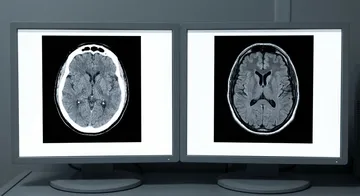
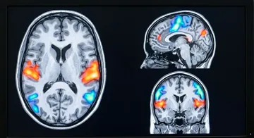
On contrast-enhanced brain MRI, meningiomas are typically observed as homogeneous, intensely contrast-enhancing masses showing a broad-based contact with the dura. The dural tail sign is a highly specific finding for meningioma.5,6 CT provides additional information in evaluating calcification and bone invasion.
Treatment Approach
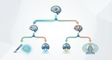
The treatment decision depends on the tumour's size, growth rate, location, the patient's age, and symptoms. Small, asymptomatic, and non-growing meningiomas can be followed up with serial MRIs (watch and wait strategy).
Surgical resection is the standard treatment for symptomatic or growing meningiomas.7,8 Surgical success is evaluated according to the Simpson classification: Grade I (complete removal of the tumour along with the involved dura and bone) has the lowest recurrence rate, while Grade V (decompression only) has the highest recurrence rate.9
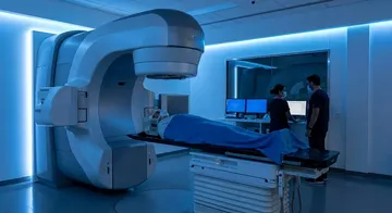
Stereotactic radiosurgery (Gamma Knife) is an effective alternative for small and surgically difficult-to-access meningiomas or for postoperative residual tumours.10,11
→ Next page: S13 — Meningioma Grade Classification and Prognosis
Kaynaklar
- Ostrom QT et al. Neuro Oncol. 2023. PMID 37793125
- Amoo M et al. Neurooncol Adv. 2023. PMID 37287581
- Telugu RB et al. J Cancer Res Ther. 2020. PMID 33342817
- Pikis S et al. Neurosurg Clin N Am. 2023. PMID 37210134
- Toda Y et al. Spine Surg Relat Res. 2025. PMID 39935985
- Rokni-Yazdi H et al. Eur J Radiol. 2006. PMID 16675180
- Goldbrunner R et al. Neuro Oncol. 2021. PMID 34181733
- Nassiri F et al. Nature. 2021. PMID 34433969
- Gallagher MJ et al. Clin Neurol Neurosurg. 2016. PMID 26780494
- Khaboushan AS et al. Acta Neurochir (Wien). 2024. PMID 38980438
- Hafez RF et al. Acta Neurochir (Wien). 2015. PMID 26231629
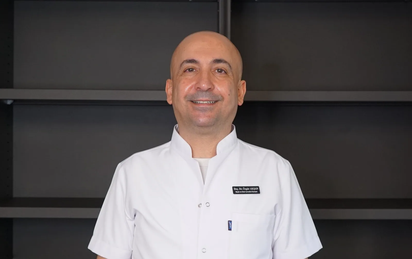
Doç. Dr. Özgür Akşan
Beyin ve Sinir Cerrahisi Uzmanı
Beyin ve Sinir · Sorumlu Yazı İşleri Müdürü ve Editör · Künye