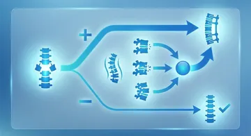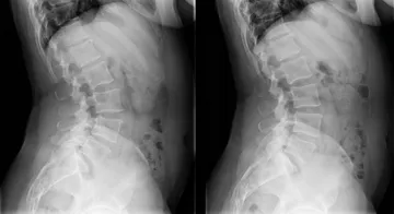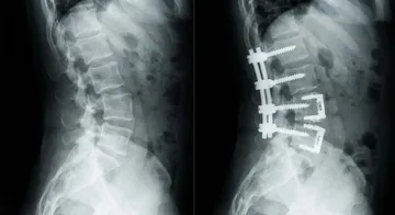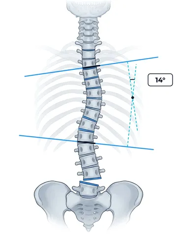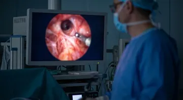
The main goal in spinal cord surgery: to remove the tumour as completely as possible and to preserve neurological function. This balance is extremely delicate — it requires millimetre-level precision.
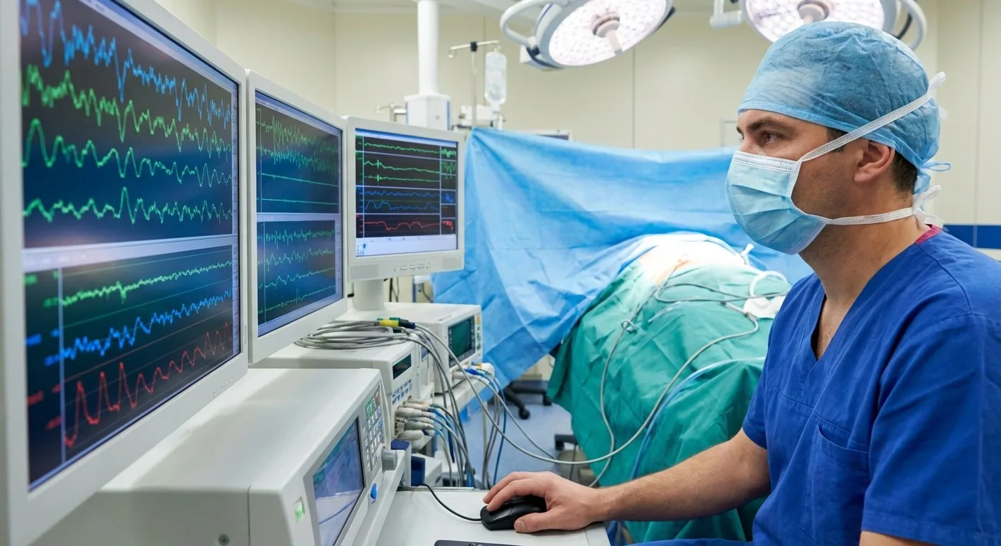
Intraoperative Neuromonitoring (IONM)
Throughout the surgery, the patient's nerve functions are monitored in real time:
- SEP (Somatosensory Evoked Potential): Monitors sensory pathways
- MEP (Motor Evoked Potential): Monitors motor pathways
- EMG: Monitors nerve root function
Technical Steps
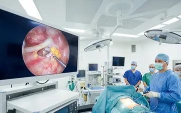
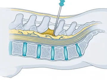
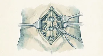
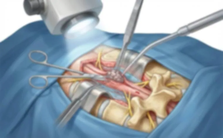
Posterior approach: The most commonly used route. The canal is opened with a laminectomy or laminoplasty. The dura is opened to reach the tumour.
In intramedullary tumours: A myelotomy is performed from the posterior median sulcus or the dorsal root entry zone. The tumour is removed using an ultrasonic aspirator (CUSA) and microsurgical techniques. The tumour-spinal cord interface is carefully determined.
In extramedullary tumours: The tumour has usually pushed the spinal cord to the side. After removal from within the capsule (intracapsular debulking), the capsule is separated from the surrounding tissues.
Laminoplasty: Especially in children, the lamina is replaced to preserve the bone structure and prevent the development of deformity.
In extramedullary tumours (meningioma, schwannoma), the total removal rate is over 90%; recurrence is rare after total removal, but it can rise above 5% in long-term follow-up.1,2
Outcomes
In extramedullary tumours (meningioma, schwannoma), the total removal rate is over 90%; recurrence is rare after total removal, but it can rise above 5% in long-term follow-up. In intramedullary ependymomas, total removal (resection) can be achieved when a dissection plane is found between the tumour and the spinal cord. In astrocytomas, subtotal removal is more common due to diffuse borders.3,4
→ Next page: S35 — Minimally Invasive Spine Surgery
Kaynaklar
- El-Hajj VG et al. Cancers (Basel). 2022. PMID 36551706
- He Z et al. Asian J Neurosurg. 2022. PMID 35873837
- Butenschoen VM et al. J Neurooncol. 2021. PMID 33094355
- Knafo S et al. Rev Neurol (Paris). 2021. PMID 33066996
Doç. Dr. Özgür Akşan
Beyin ve Sinir Cerrahisi Uzmanı
Beyin ve Sinir · Sorumlu Yazı İşleri Müdürü ve Editör · Künye