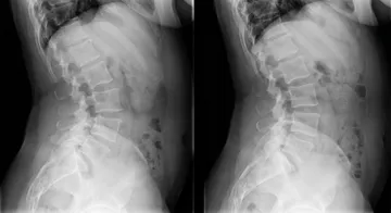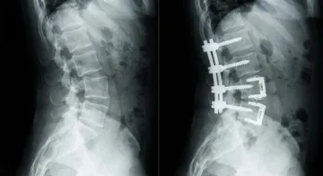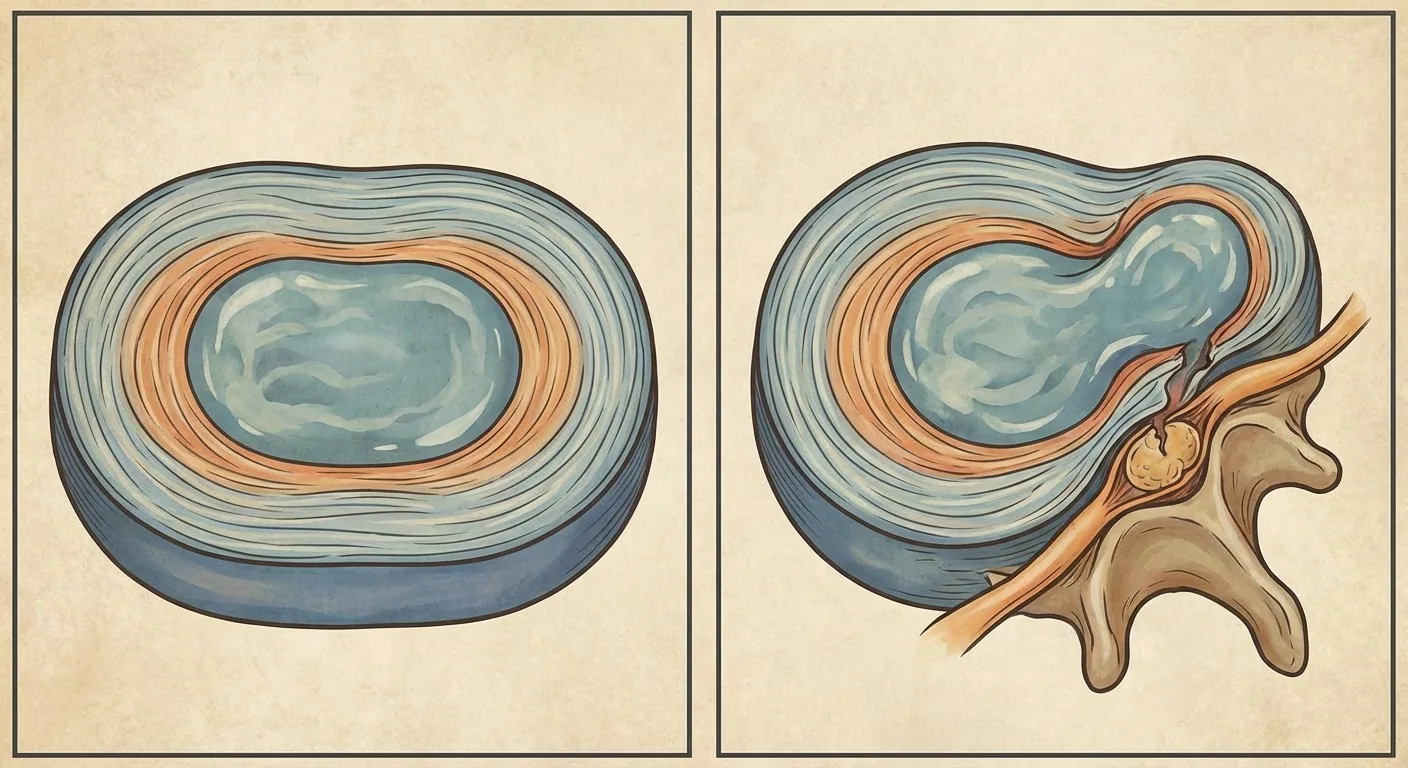
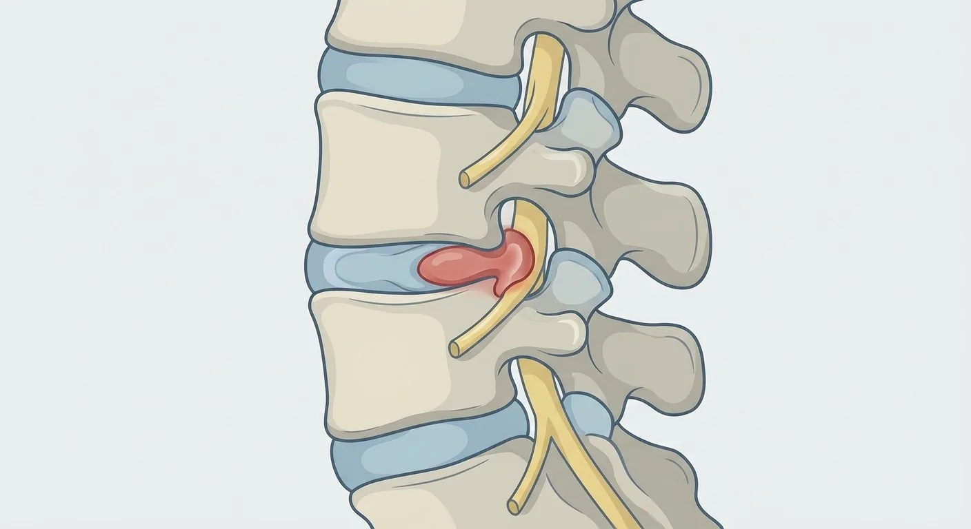
A herniated disc is a spinal condition that affects approximately 1-2.5 percent of the population and is most commonly seen between the ages of 30-50.1 It occurs when one of the discs between the vertebrae tears, causing the inner core material (nucleus pulposus; Latin nucleus pulposus) to leak out and press on a nerve root.
Structure of the Spine
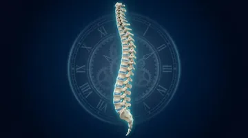
The lower back region (lumbar spine) consists of five vertebrae (L1-L5) and the discs between them. Each disc has an outer tough fibrous ring (annulus fibrosus) and an inner gel-like core (nucleus pulposus). The discs act as shock absorbers during spinal movements. Ageing, trauma, or repetitive microtraumas can cause a tear in the annulus fibrosus, paving the way for herniation.
Types of Herniation
Disc pathologies are classified into four stages. Bulging: the disc margins expand but the annulus is intact. Protrusion: the annulus begins to tear, and the nucleus partially bulges out. Extrusion: the nucleus completely breaches the annulus. Sequestration: the detached fragment becomes free-floating.
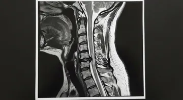
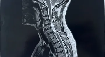
Lumbar MRI is the gold standard imaging method that shows the herniation and its relationship with the nerve.
Symptoms
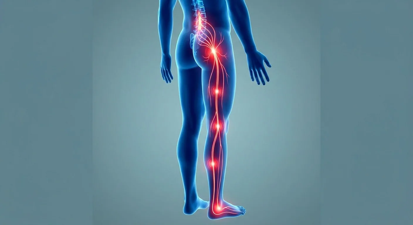
Low back pain is the most common initial symptom. Sciatica (sciatic pain) — pain radiating from the buttock down the leg, sometimes all the way to the foot — is the most typical symptom of a herniated disc. The pain usually increases with coughing, sneezing, and straining. Numbness, tingling, and loss of strength in the leg are signs of nerve root damage.
Cauda equina syndrome is a condition that requires emergency surgery: loss of strength in both legs, loss of bowel and bladder control, and numbness in the saddle area (perineum).2 If these symptoms are present, you should immediately go to the emergency room.
Diagnosis
History and physical examination form the basis of diagnosis. The straight leg raise test (Lasegue) and neurological examination evaluate nerve root compression. Lumbar MRI is the gold standard imaging method; it shows the level and type of the herniation, as well as its relationship with the nerve root.3,4 EMG (electromyography) objectively measures the degree of nerve damage.
Treatment Approach
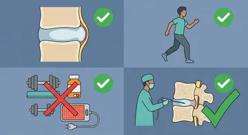
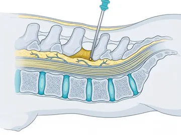
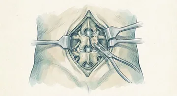
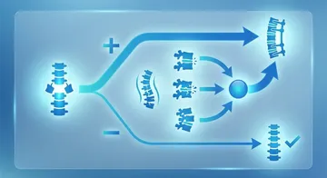
The vast majority of herniated disc patients (80-90 percent) recover with conservative treatment. Medication, physical therapy, lifestyle modifications, and, when necessary, epidural injections are the components of conservative treatment. Surgery is considered for patients who do not respond to 6-8 weeks of conservative treatment, develop progressive neurological deficit, or have cauda equina syndrome.5,6
→ Next page: S20 — Microdiscectomy and Endoscopic Surgery
Kaynaklar
- Benditz A et al. Orthopadie (Heidelb). 2026. PMID 42384175
- Najjar E et al. Spine J. 2026. PMID 41991105
- Pojskic M et al. World Neurosurg X. 2024. PMID 38440379
- Wang Y et al. Sci Rep. 2025. PMID 40999029
- Kögl N et al. Dtsch Arztebl Int. 2024. PMID 38835174
- Yoon WW et al. EFORT Open Rev. 2021. PMID 34267943
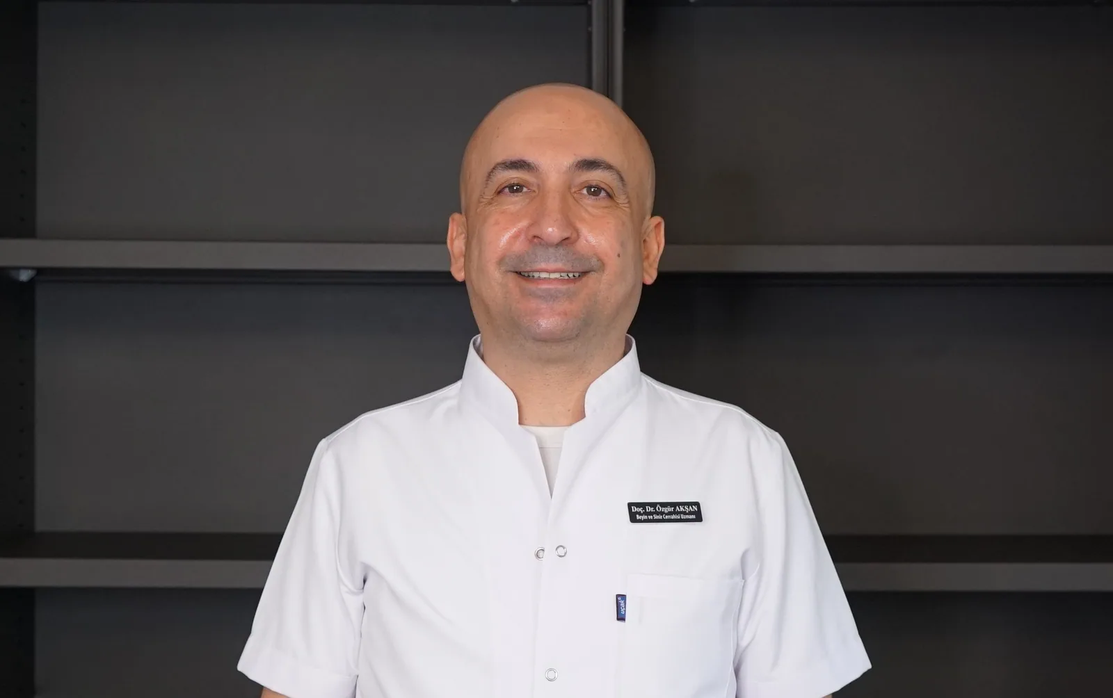
Doç. Dr. Özgür Akşan
Beyin ve Sinir Cerrahisi Uzmanı
Beyin ve Sinir · Sorumlu Yazı İşleri Müdürü ve Editör · Künye